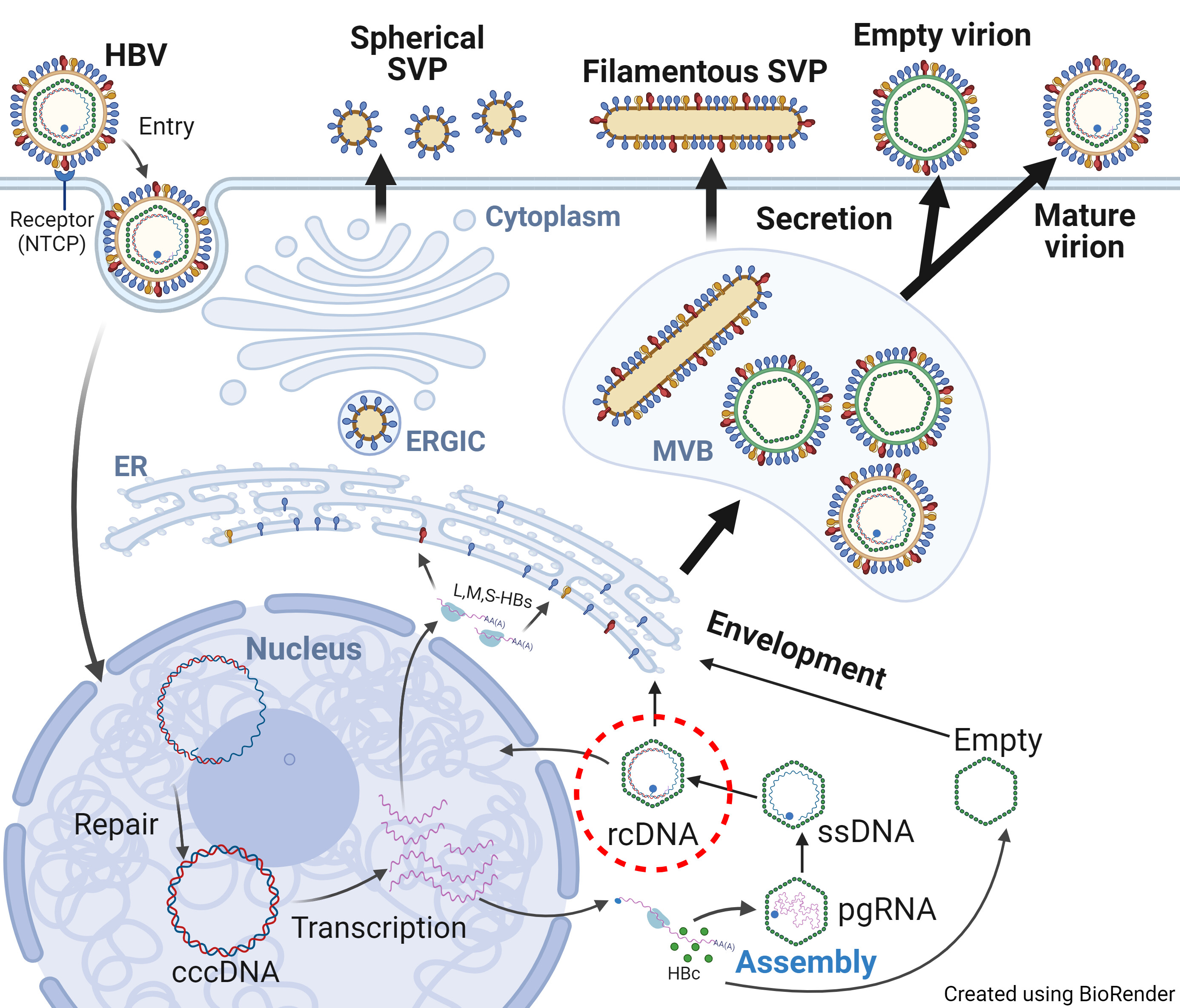
Hello everyone! My name is Joseph Wang, and I am excited to join this forum and see so many discussions about HBV research. I am part of the research team at Penn State College of Medicine, where we are passionate about solving important questions related to human diseases. Our lab focuses on understanding the different structures of the hepatitis B virus (HBV) throughout its life cycle. In simple terms, we want to see how HBV looks when it infects human cells, changes inside the cells, breaks down to deliver its genetic material, and how new viruses are assembled and released to infect new cells (Figure 1).
You might think we already know all this because HBV has been studied for a long time, but that’s only partly true. In the past, scientists have used mutated HBV proteins produced in bacteria to study the virus’s structure. However, these studies don’t always give us the full picture. The virus parts they studied might be missing some important information, such as lacking proper modifications to the protein that occur in human cells or even containing bacterial DNA instead of viral DNA.
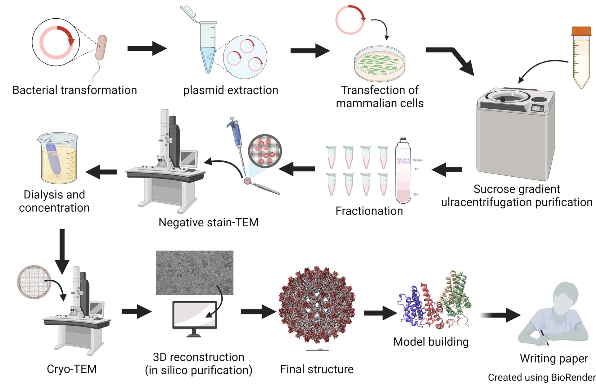
In our lab, we study the native structure of HBV by putting an HBV replicon (a gene that makes all HBV proteins except for its envelope proteins) into liver cancer cells or by collecting HBV particles from human patients or experimental animals. After purifying these particles, we freeze them very quickly using liquid ethane, which is extremely cold. This quick freezing preserves the virus in its natural state. We then use a transmission electron microscope, called cryo-EM, to take thousands of images of the virus and use software to piece together a detailed structure (Figure 2, image credit: Emily Bianchini).
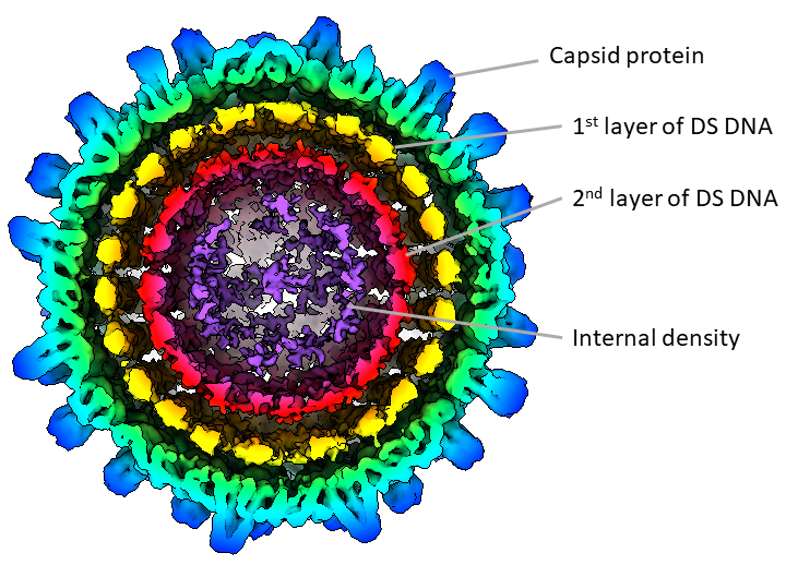
One of the structures that we are working on is the mature HBV DNA capsid (Figure 3). By using the HBV replicon, we produced HBV particles that have different types of genetic material (RNA, single-stranded DNA, and partially double-stranded DNA). Using computational methods, we could select the particles with double-stranded DNA. We discovered that the mature DNA particle has multiple layers of density inside, organized into rings like a compressed spring, suggesting internal pressure. We also found some masses inside the particle, which we are trying to identify - they might be reverse transcriptase or other host proteins. We are working to get a clearer picture of this structure to understand it better.
So, why is it important to “see” the structure? There is an old saying: “seeing is believing.” By understanding the detailed structure of the mature HBV capsid, we can compare it with other HBV structures to learn how the virus changes during its life cycle. This information can also help us understand how drugs interact with the virus, which could lead to better treatments.
Thank you for taking the time to read about our research. I am happy to answer any questions you may have! ![]()